
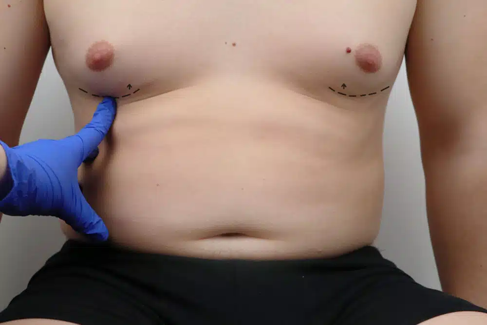
Male chest contour correction with glandular resection and liposculpting for a flat, athletic thoracic profile.
Gynecomastia Correction: Clinical Management of Male Mammary Morphology
The surgical correction of excessive male breast tissue, clinically identified as Gynecomastia, represents a specialized intervention designed to restore the structural proportions of the male chest. This condition often arises from a hormonal imbalance between estrogen and androgen, leading to the proliferation of both glandular (fibrous) and adipose (fatty) tissues. The primary objective is the systematic removal of this excess volume to achieve a more contoured and athletic thoracic profile. By meticulously managing the relationship between the skin envelope and the underlying pectoral muscles, surgeons can eliminate the appearance of “female-like” breasts and enhance the patient’s physical confidence and comfort.
Gynecomastia Correction procedures integrate advanced liposculpting methodologies with precise parenchymal resection to address the benign proliferation of male mammary tissue. By strategically modulating localized adipose deposits and eliminating redundant glandular structures, these interventions restore a flat, firm, and anatomically masculine thoracic silhouette. Each clinical strategy is meticulously tailored to maintain neurovascular integrity and optimize dermal adherence while achieving a resilient, symmetrical, and harmoniously contoured aesthetic transformation
1. Differential Diagnosis and Tissue Composition
The technical execution of Gynecomastia Correction is predicated on a thorough preoperative assessment of the tissue composition. Clinical gynecomastia is categorized into three types: Glandular (predominantly firm breast tissue), Fatty (Pseudogynecomastia), and Multicomponent (a mixture of both). Identifying the ratio of fibrous gland to subcutaneous fat is critical for selecting the appropriate surgical modality. In cases where glandular tissue is prominent, a direct surgical excision is required, whereas fatty tissue can often be effectively addressed through specialized suction-assisted techniques.
Liposuction-Assisted Contouring and VASER Technology
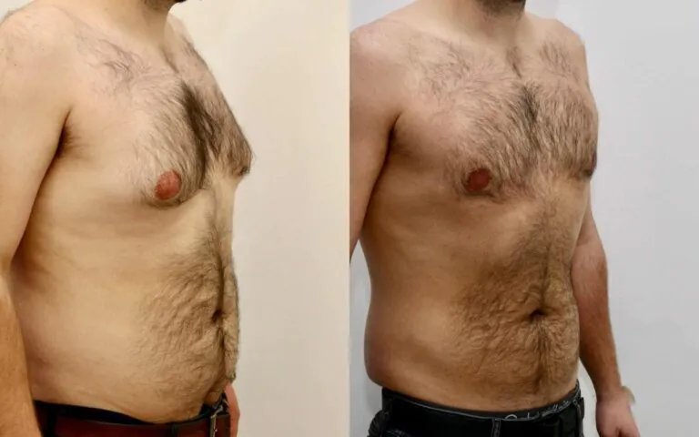
For the majority of patients, the first phase of the procedure involves Liposuction to debulk the surrounding adipose tissue and define the borders of the pectoral muscle. The integration of VASER (Ultrasonic-Assisted) Liposuction has revolutionized this process by utilizing ultrasonic energy to emulsify fat cells while preserving the integrity of blood vessels and nerves. This advanced technology allows for more aggressive contouring of the chest wall and promotes superior skin contraction, ensuring that the dermal envelope adheres smoothly to the new underlying framework.
Subcutaneous Mastectomy and Glandular Resection
In cases where a firm, subareolar mass persists after liposuction, a Subcutaneous Mastectomy is performed through a discreet periareolar incision. This involves the direct surgical resection of the hypertrophied mammary gland. A critical priority during this phase is the preservation of a small “button” of tissue beneath the nipple-areolar complex (NAC) to prevent a “crater” deformity or nipple inversion. By achieving a smooth transition between the resected zone and the surrounding subcutaneous fat, surgeons ensure a natural and symmetrical thoracic profile.
Dermal Envelope Management and Skin Redundancy
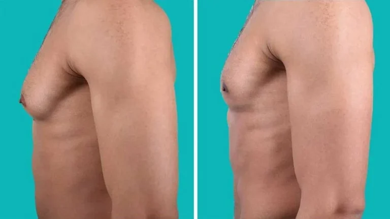
In patients with significant skin laxity (Grade III or IV Gynecomastia), the resection of redundant skin may be necessary to achieve an optimal result. This may involve periareolar or extended skin excision techniques similar to a mastopexy.
The goal is to reposition the NAC to a more superior and anatomically correct coordinate while tightening the integumentary envelope. By strategically placing incisions within natural anatomical boundaries, clinicians minimize visible scarring and ensure that the chest appears firm and well-proportioned.
Postoperative Stabilization and Pectoral Integration
The recovery phase following Gynecomastia Correction is governed by the management of interstitial edema and the promotion of tissue adherence. Patients are integrated into a progressive protocol that includes the use of medical-grade compression vests for 4 to 6 weeks.
This external support is essential to minimize seroma formation and to help the skin settle into its new masculine contours. Continuous clinical monitoring ensures that the histological remodeling results in a stable, refined, and enduring outcome, adhering to the principles of anatomical permanence and structural excellence.